
Thermal or infrared imaging fills the gap in clinical diagnosis; X-ray, CT scans, ultrasound, and MRI are all tests of anatomy or structure. Thermal imaging is unique in its capability to show physiological or functional changes and metabolic processes. It has proven useful as a complementary procedure to other diagnostic efforts. Thermal Imaging uses a patented computerized camera that detects radiated heat and measures the physiological activity in the body. The procedure requires no physical contact or liquids to drink. Thermal patterns depict tissue, muscle, joint and organ function.
Since it is non-invasive and incredibly sensitive, this technology offers graphic mapping and display of all skin surface temperatures. Thermal imaging allows us to diagnose, evaluate, monitor, and documents a wide range of injuries and conditions. And it can graphically display the biased feelings of pain by accurately display the changes in the skin surface temperature. Thermal imaging can help diagnose treat painful conditions like Fibromyalgia, Rheumatoid Arthritis, and Complex Regional Pain Syndrome (CRPS/RSD). It can show a combined effect of the autonomic nervous and the vascular system, even down to the capillary level.
Thermal imaging can help us get to the root cause of your pain, and design a treatment plan that delivers results!
Here are some examples of thermal imaging performed in our clinic:

Head image of patient suffering headache pain.
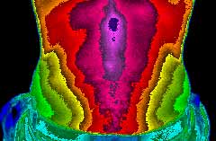
Lower abdomen image of patient, evaluating for organ-referred pain.
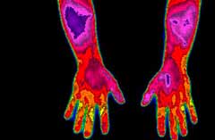
Hand images of patient suffering from forearm pain.